Chung-Shih Chen1,2,
Mei-Lin Wang2,3,
Rosa Huang Lin4,
Shih-Pin Chen5,6,7,
Shih-Pin Chen5,6,7,
Tsong-Ming Lu5,6,8,
Wei-Yu Tsai9,
Chien-Fu Huang10,
Chi-Chiang Yang9,11,
Yew-Min Tzeng1,12,13 ![]() ,
,
For correspondence:- Yew-Min Tzeng Email: cyang@csmu.edu.tw Tel:+886424730022
Received: 8 October 2015 Accepted: 15 February 2016 Published: 31 March 2016
Citation: Chen C, Wang M, Lin RH, Chen S, Chen S, Lu T, et al. Anti-fatigue effect of aqueous extract of Anisomeles indica (L) Kuntze in mice. Trop J Pharm Res 2016; 15(3):489-495 doi: 10.4314/tjpr.v15i3.9
© 2016 The authors.
This is an Open Access article that uses a funding model which does not charge readers or their institutions for access and distributed under the terms of the Creative Commons Attribution License (http://creativecommons.org/licenses/by/4.0) and the Budapest Open Access Initiative (http://www.budapestopenaccessinitiative.org/read), which permit unrestricted use, distribution, and reproduction in any medium, provided the original work is properly credited..
Purpose: To determine the anti-fatigue effect of Anisomeles indica (L.) Kuntze, an herb traditionally used for health improvement in Taiwan.
Methods: Three groups (n = 10 per group) of Balb/c female mice were administered A. indica aqueous extract orally for 28 days at 125 (low dose A. indica, LA), 250 (medium dose A. indica, MA), and 500 (high dose A. indica, HA) mg/kg/day, respectively, while a control group received distilled water. After 28 days, a forced swimming test was performed, and biochemical parameters including plasma triglyceride (TG), glucose, lactate and ammonia levels related to fatigue were examined.
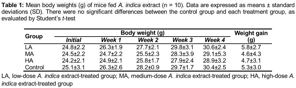
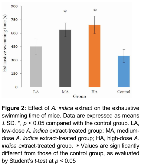
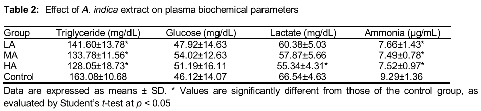
Results: No mice died during the study period. Physical examinations did not reveal any treatment-related adverse effects after dosing, in terms of food and water consumption. Moreover, no obvious peptic ulcers, haemorrhage, or pathological changes in the liver or kidney were observed in A. indica-treated mice, and there were no significant differences in body weight between the control and treatment groups (p > 0.05). Mice treated with A. indica extract in the MA and HA groups showed significantly prolonged exhaustive swimming time (p < 0.05), increased hepatic glycogen and muscle glycogen levels (p < 0.05), and decreased triglyceride and plasma ammonia levels (p < 0.05) in a dose-dependent manner, compared with the controls. However, plasma glucose and lactic acid levels were not significantly changed (p > 0.05).
Conclusion: These results provide the first in vivo evidence supporting the anti-fatigue claims associated with A. indica treatment, indicating that this traditional herb may be of therapeutic use as an ergogenic and anti-fatigue agent
Introduction
Fatigue, the phenomenon of decreased efficiency following a period of continuous study or work, may be subdivided into mental and physical fatigue. Physical fatigue manifests mainly as physical deteriorations of muscle tone and exercise tolerance, due to an accumulation of lactic acid and other metabolites [1]. Regular exercise combined with a balanced diet may be the most effective strategy through with to maintain or promote good health [2]. Several studies have shown that exogenous antioxidants can reduce exercise-induced oxidative stress [3]. For the sake of convenience, more and more people are choosing to take complementary and alternative medicines to eliminate fatigue-related metabolites and improve athletic ability.
Previous studies have shown that herbal medicines can improve various parameters of immune function [4,5] and may provide effective complementary support for cancer patients [6,7]. Research on specific nutrients or herbal supplements is required to identify agents that reduce metabolite production and/or improve energy utilization.
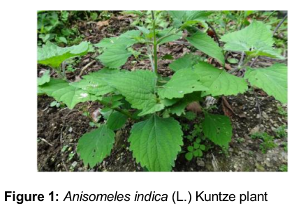
Anisomeles indica (family Lamiaceae) () is an erect camphor-scented perennial woody shrub that grows in the wild in Southeast Asia. Anisomeles indica Kuntze, which is commonly known as “yu-zhen-tsao”, is the only species of Anisomeles found in Taiwan [8,9]. It is used in folk medicine for the treatment of diverse conditions, such as inflammatory disease, liver disease, intestinal infections, abdominal pain and immune system deficiencies [10]. Although A. indica has been used for health improvement in Taiwan, its anti-fatigue effects have not yet been reported.
In the present study, aqueous extract of A. indica was administered to three groups of mice, and exhaustive swimming times and relevant physiological variables were measured to determine the in vivo anti-fatigue effects of the extract.
Methods
Preparation of medicinal plant extract
Whole plants of A. indica were collected from Yuli, Hualien County in Eastern Taiwan, and a botanically identified voucher specimen (YMT-09–02) was deposited in the herbarium of the Institute of Biochemical Sciences and Technology, Chaoyang University of Technology, Taiwan. A. indica extract was prepared according to standard extraction and concentration procedures. Briefly, whole plants (1.4 kg) were air-dried at room temperature and decocted in water (4 × 5 L) for 1 h. The resulting extract was filtered and concentrated under reduced pressure, producing a dark brown syrup (36.5 g, 2.6 %, w/w), which was stored in the freezer. The moisture content of the extract was determined as follows: 1 g final extract was placed in an oven at 60 – 65 °C for 72 h and then weighed; the weight lost via drying was used as an indicator of the moisture content. The final extract contained 14 % water. This extract was dissolved or suspended in distilled water prior to oral administration to the mice.
Animals and grouping
The Institutional Guidelines for Animal Experiments (Animal Center, Chung Shan Medical University) were followed for animal care and use in the experiments (approval no. 1282). The mice used in this study were 8-week-old specific pathogen-free (SPF) Balb/c female mice, purchased from the National Laboratory Animal Center, Taipei, Taiwan. These animals were raised in the Animal Center of Chung Shan Medical University at a temperature of 25 ± 1 °C and under 55 ± 5 % relative humidity, with 12 h of daily light exposure.
After 1 week of acclimatization, the animals were divided randomly into four groups (n = 10): a) control group (administered distilled water with no A. indica extract), b) low-dose A. indica extract (125 mg/kg)-treated group (LA), c) medium-dose A. indica extract (250 mg/kg)-treated group (MA), and d) high-dose A. indica extract (500 mg/kg)-treated group (HA). The mice in each group were orally administered the extract in 1 ml volumes daily for 28 days.
Forced swimming test
The forced swimming capacity test was employed in this study to evaluate the effects of medicinal plant extracts on exercise durability in mice. Swimming is commonly accepted as an experimental exercise model [11]. After 28 days of treatment, 10 mice from each group were subjected to the forced swimming test. The procedure used was as described previously [2], with slight modifications. Thirty minutes after the final treatment, the mice were placed individually into an acrylic plastic tank (50 × 50 × 40 cm) containing water at a 30 cm depth and maintained at 37 ± 1 °C. A tin wire (5 % body weight) was loaded onto the tail root of each mouse. The mice were considered to be exhausted when they failed to rise to the surface of the water to breathe within a 10 s period, at which point the total swimming time was recorded immediately.
Analysis of biochemical parameters of blood
After anesthetization with ether, whole blood samples were collected from mice in heparinized tubes using a heart puncture technique. Plasma was isolated by centrifugation at 900 × g, 4 °C for 10 min and stored at −70 °C in a deep-freezer. The plasma triglyceride (TG), glucose, lactate and ammonia levels were analysed using commercial kits (Sigma-Aldrich Chem. Co., St. Louis, MO, USA).
Analysis of tissue glycogen contents
Immediately after blood collection, the liver and gastrocnemius muscle were isolated quickly, frozen in liquid nitrogen, and stored at −70 °C prior to analysis of glycogen content. The glycogen content was measured spectrophotometrically according to methods described previously [12]. The liver and gastrocnemius muscle samples (1 g) were hydrolysed in 30 % KOH at 100 °C for 30 min; 1.5 mL anhydrous ethanol was then added to the vials, which were centrifuged at 4000 × g for 15 min. The supernatant was discarded. Then, 0.5 mL distilled water and 1 mL 0.2 % anthrone (0.2 g anthrone in 100 ml 98 % H2SO4 [g/ml], prepared freshly over 1 h) were added to the vials, which were then placed in a boiling water bath for 20 min. The absorbance of the solution at 620 nm was determined using a spectrophotometer (V-530, Jasco Co., Japan).
Statistical analysis
The results are expressed as mean ± standard deviation (SD). The statistical significance of the differences between the control and each treatment group was determined using the Student’s t-test, and p < 0.05 was considered statistically different. All the analyses were performed using SPSS 15 (SPSS Inc., Chicago, IL, USA).
Results
Effect of A. indica treatment on gross pathology and body weight
No mice died during the experimental period. Moreover, there were no obvious signs of peptic ulcers, haemorrhage, or pathological changes in the liver or kidney observed in any of the A. indica-treated mice (data not shown). To assess the effects of the treatments on toxicity, body weight and food and water consumption were monitored throughout the study. A. indica extract, at doses of 125, 250, or 500 mg/kg daily, was not found to be associated with any apparent toxicity, as evaluated by monitoring food and water consumption (data not shown). Body weight was recorded before the experiment (initial) and after 4 weeks, and the weight differences were analysed statistically. There was no significant difference between the control group and each treatment group. The changes in body weight during the experimental period are shown in .
Effect of A. indica treatment on forced swimming capacity
The swimming times to exhaustion of the LA, MA, HA, and control groups were 452.3 ± 85.2, 638.5 ± 77.7, 692.5 ± 96.8, and 346.5 ± 72.1 s, respectively (). There were obvious differences in the swimming time to exhaustion between the control group and each treatment group. The swimming times to exhaustion of the A. indica extract-treated mice in the MA, and HA groups were statistically greater than that of the control group. These effects were dose-dependent (p < 0.05).
Effect of A. indica treatment on plasma biochemistry
The results of the biochemical analyses of the plasma samples from each group are shown in . The plasma TG levels were lower in all of the A. indica extract-treated groups, compared with the control group (p < 0.05). However, the plasma glucose levels in the A. indica extract-treated groups were slightly higher than those of the control group; however, no statistically significant differences were observed. Moreover, there was no statistically significant difference in the plasma lactate level between the A. indica extract-treated groups and the control group, except for the HA group, which exhibited lower lactate levels that those of the control group (p < 0.05). Nevertheless, the plasma ammonia levels were significantly lower in all of the A. indica extract-treated groups, compared with the control group (p < 0.05).
Effect of A. indica treatment on hepatic and gastrocnemius muscle glycogen levels
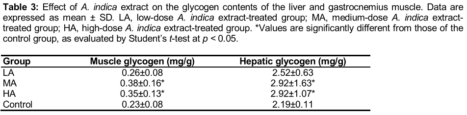
As shown in , the hepatic and muscle glycogen levels in mice of the MA and HA groups were increased significantly, compared with the control group (p < 0.05).
Discussion
In this study, we demonstrated that mice treated with A. indica aqueous extract (125, 250, and 500 mg/kg) for 28 days had longer exhaustive swimming times than those of mice treated with distilled water. Prolonged, high-intensity exercise leads to an inadequate oxygen supply at the physiological level. To maintain physical functioning, the body primarily metabolizes glucose in a process dependent on the glycolytic pathway and results in significant consumption of glycogen and accumulation of fatigue-inducing metabolites, including lactic acid [1].
The forced swimming test is frequently used to evaluate the anti-fatigue effects of potential therapeutic compounds, because it is highly effective in evaluating the endurance capacity of mice, with highly reproducible results. The length of the exhaustive swimming time indicates the degree of fatigue [12,13]. During prolonged exercise, the development of fatigue is closely related to the depletion of glycogen stores, both in liver tissue and in skeletal muscle [14,15]. Glycogen stores, which are an important energy source during exercise, complement the uptake of glucose from blood, and are important in maintaining blood glucose levels within the physiological range [16]. Fatigue occurs when glycogen stores are nearly exhausted. Glycogen is therefore a sensitive indicator of fatigue, and increased glycogen storage is associated with enhanced endurance and locomotory capacities [17].
In the present study, biochemical analyses revealed that the liver and muscle glycogen concentrations of the mice in the A. indica-treated groups were significantly increased compared with those in the control group. The plasma TG concentration was decreased in the treatment groups relative to the control group. These results suggests that A. indica extract may increase fat utilization in mice during swimming, and the anti-fatigue effects of A. indica extract might be related to an improvement in the activation of energy conversion.
Biochemical variables, including lactate, ammonia, glucose, and creatine kinase, are important indicators of muscle fatigue after exercise [18]. Ammonia, which is a metabolite of proteins and amino acids, was linked to fatigue as early as 1922 [19]. The increase in ammonia in response to exercise is managed through the use of amino acids or carbohydrates that interfere with ammonia metabolism [20]. The increase in ammonia levels is related to both peripheral and central fatigue during exercise. Since excess ammonia has a toxic effect on the central nervous system, exercise-induced ammonia accumulation may contribute to the induction of central fatigue. Blood lactate levels were determined primarily as an index of anaerobic metabolism during swimming [21]. Increased lactate results in a further reduction in blood pH, which may induce various biochemical and physiological side effects, including glycolysis and the release of phosphofructokinase and calcium ions, through muscular contraction [22]. After administration of A. indica extract to mice for 28 days, plasma ammonia levels were significantly lower after the swim test in the treated groups, compared with the control group. However, lactic acid levels were not significantly changed, although non-significant decreases were observed in the A. indica extract-treated groups. Taken together, these data suggest that administration of A. indica extract can alleviate fatigue in mice during exercise.
Previously, it has been reported by our and other research groups that A. indica extracts and isolated constituents inhibit inflammatory mediators and tumor cell proliferation [23-25]. Furthermore, the aqueous extract of A. indica has been shown to possess anti-histaminergic, anti-hyperalgesic and analgesic activities [26, 27]. In addition, ethanol extract of A. indica exhibits anti-bacterial activity [28], and methanol extract displays antioxidant capacities [10]. Moreover, the constituents of A. indica have potent in vitro inhibitory effects on the production of the inflammatory mediators nitric oxide, tumor necrosis factor (TNF-α), and interleukin-12 (IL-12) [25].
Interestingly, it was reported that mice treated with coenzyme Q10 showed a significantly prolonged exhaustive swimming time, increased liver glycogen contents, and decreased serum urea nitrogen levels, compared with the control animals; however, lactic acid levels were not significantly changed [29]. The anti-fatigue effects of A. indica are quite similar to those of coenzyme Q10, which has been shown to be a promising anti-fatigue agent.
Conclusion
The data presented here indicate that A. indica extract exhibits anti-fatigue effects by decreasing plasma TG and ammonia levels and increasing liver and muscle glycogen deposition, thereby increasing the exercise performance of the mice in this study. To achieve good efficacy and safety, a daily dose of A. indica extract ranging from 250 to 500 mg/kg is suggested. Although the bioactive component(s) in the extract and the detailed anti-fatigue mechanism remain to be elucidated, this study provides the first in vivo evidence to support claims of the anti-fatigue effect of A. indica treatment. A. indica may, therefore, be a useful ergogenic and anti-fatigue therapeutic agent.
Declarations
Acknowledgement
References
Archives


News Updates